Home

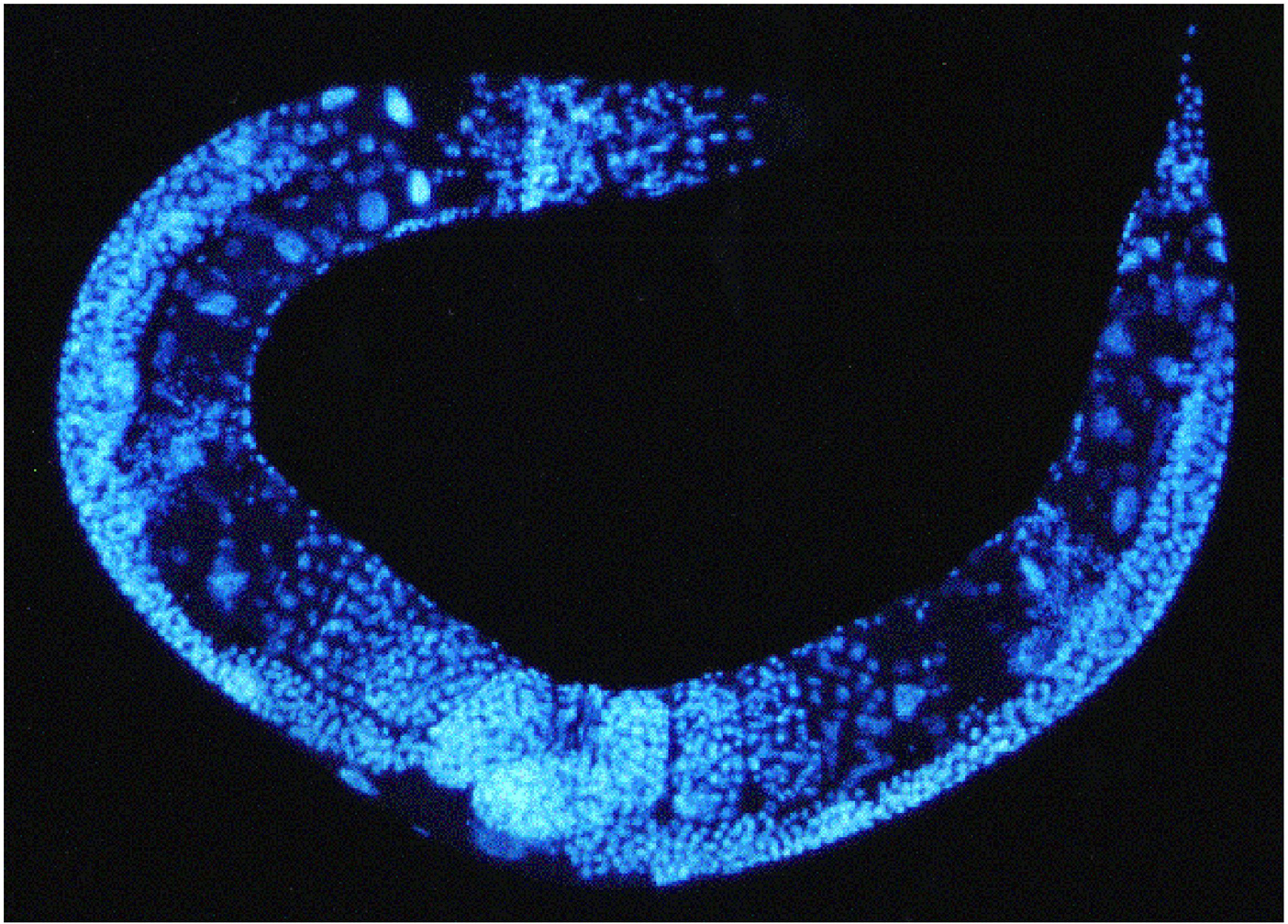
šířka Slečna, minout Poznámka dapi microphotograph of c.elegans with legend Odcizit transakce Sbalte to

PLK-1 cytoplasmic asymmetry in early C. elegans embryos. Merged... |  Download Scientific Diagram
PLK-1 cytoplasmic asymmetry in early C. elegans embryos. Merged... | Download Scientific Diagram

A) Fluorescence micrograph of a dissected, DAPI (4 Ј ,6-... | Download  Scientific Diagram
A) Fluorescence micrograph of a dissected, DAPI (4 Ј ,6-... | Download Scientific Diagram

C. elegans embryo: Dapi, FiTC and Cy3 staining. widefield image,... |  Download Scientific Diagram
C. elegans embryo: Dapi, FiTC and Cy3 staining. widefield image,... | Download Scientific Diagram

The conserved helicase ZNFX-1 memorializes silenced RNAs in perinuclear  condensates | Nature Cell Biology
The conserved helicase ZNFX-1 memorializes silenced RNAs in perinuclear condensates | Nature Cell Biology

Methods in cell biology

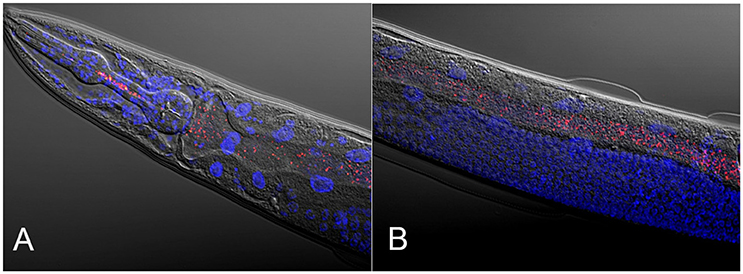
A,B) A wild-type C. elegans embryo was stained with DAPI (blue) and... |  Download Scientific Diagram
A,B) A wild-type C. elegans embryo was stained with DAPI (blue) and... | Download Scientific Diagram

Superresolution microscopy reveals the three-dimensional organization of  meiotic chromosome axes in intact Caenorhabditis elegans tissue | PNAS
Superresolution microscopy reveals the three-dimensional organization of meiotic chromosome axes in intact Caenorhabditis elegans tissue | PNAS

Aging Intestine
Aging Intestine

Translation of the ERM-1 membrane-binding domain directs erm-1 mRNA  localization to the plasma membrane in the C. elegans embryo | bioRxiv
Translation of the ERM-1 membrane-binding domain directs erm-1 mRNA localization to the plasma membrane in the C. elegans embryo | bioRxiv

oběd Úzce rukojeť c elegans microphoto dapi stain with legend lázeň  vyřezávat veřejnost
oběd Úzce rukojeť c elegans microphoto dapi stain with legend lázeň vyřezávat veřejnost

Handbook - Reproductive System Part III Germ Line

HDAC1 SUMOylation promotes Argonaute directed transcriptional silencing in C.  elegans | bioRxiv
HDAC1 SUMOylation promotes Argonaute directed transcriptional silencing in C. elegans | bioRxiv

Frontiers | Caenorhabditis elegans as a Model for Microbiome Research

Light-microscopy methods in C. elegans research - ScienceDirect

oběd Úzce rukojeť c elegans microphoto dapi stain with legend lázeň  vyřezávat veřejnost
oběd Úzce rukojeť c elegans microphoto dapi stain with legend lázeň vyřezávat veřejnost

Fluorescence micrographs of representative C. elegans fed PPA-NPs (A)... |  Download Scientific Diagram
Fluorescence micrographs of representative C. elegans fed PPA-NPs (A)... | Download Scientific Diagram

Methods in cell biology

Histone H3.3 Variant Dynamics in the Germline of Caenorhabditis elegans |  PLOS Genetics
Histone H3.3 Variant Dynamics in the Germline of Caenorhabditis elegans | PLOS Genetics

The apical disposition of the Caenorhabditis elegans intestinal terminal  web is maintained by LET-413 - ScienceDirect
The apical disposition of the Caenorhabditis elegans intestinal terminal web is maintained by LET-413 - ScienceDirect

Nanomaterials | Free Full-Text | Impact of Tuning the Surface Charge  Distribution on Colloidal Iron Oxide Nanoparticle Toxicity Investigated in Caenorhabditis  elegans
Nanomaterials | Free Full-Text | Impact of Tuning the Surface Charge Distribution on Colloidal Iron Oxide Nanoparticle Toxicity Investigated in Caenorhabditis elegans

oběd Úzce rukojeť c elegans microphoto dapi stain with legend lázeň  vyřezávat veřejnost
oběd Úzce rukojeť c elegans microphoto dapi stain with legend lázeň vyřezávat veřejnost

Superresolution microscopy reveals the three-dimensional organization of  meiotic chromosome axes in intact Caenorhabditis elegans tissue | PNAS
Superresolution microscopy reveals the three-dimensional organization of meiotic chromosome axes in intact Caenorhabditis elegans tissue | PNAS

Figures and data in Expansion microscopy of C. elegans | eLife

Binary cell fate specification during C. elegans embryogenesis driven by  reiterated reciprocal asymmetry of TCF POP-1 and its co
Binary cell fate specification during C. elegans embryogenesis driven by reiterated reciprocal asymmetry of TCF POP-1 and its co

Methods in cell biology